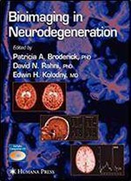
Bioimaging In Neurodegeneration (contemporary Neuroscience)
by Patricia A. Broderick /
2005 / English / PDF
14 MB Download
Bioimaging is in the forefront of medicine for the diagnosis and helps to predict the progression of AD via mild cognitive treatment of neurodegenerative disease. Conventional magnetic impairment (MCI) studies. resonance imaging (MRI) uses interactive external magnetic fields Novel neuroimaging technologies, such as neuromolecular and resonant frequencies of protons from water molecules. imaging (NMI) with a series of newly developed BRODERICK ® However, newer sequences, such as magnetization-prepared rapid PROBE sensors, directly image neurotransmitters, precursors, acquisition gradient echo (MPRAGE), are able to seek higher and metabolites in vivo, in real time and within seconds, at separate levels of anatomic resolution by allowing more rapid temporal and selective waveform potentials. NMI, which uses an imaging. Magnetic resonance spectroscopy (MRS) images electrochemical basis for detection, enables the differentiation of metabolic changes, enabling underlying pathophysiologic neurodegenerative diseases in patients who present with mesial dysfunction in neurodegeneration to be deciphered. Neuro- versus neocortical temporal lobe epilepsy. In fact, NMI has some 1 chemicals visible with proton H MRS include N-acetyl aspartate remarkable similarities to MRI insofar as there is technological (NAA), creatine/phosphocreatine (Cr), and choline (Cho) NAA dependence on electron and proton transfer, respectively, and is considered to act as an in vivo marker for neuronal loss and/or further dependence is seen in both NMI and MRI on tissue neuronal dysfunction. By extending imaging to the study of composition such as lipids.
