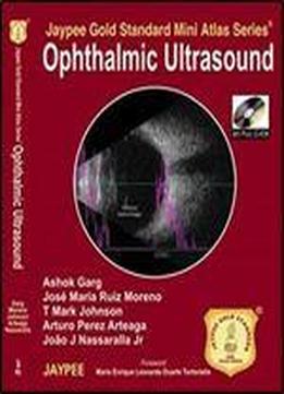
Jaypee Gold Standard Mini Atlas Series Ophthalmic Ultrasound
by Garg Ashok /
2010 / English / PDF
12.2 MB Download
The ultrasonic method is the best way to calculate the axial length and get the desired postoperative refraction before intraocular implantation. IOL power estimation formulae utilize additional preoperative biometric data of the eye, like, central anterior chamber depth (ACD), corneal diameter and the refractive error. ACD can be estimated by ultrasonography, partial coherence interferometry (PCI), scanning-slit topography and other different methods to estimate corneal power of the eye after corneal refractive surgery are refractive history, contact lens over-refraction (CLO), videokeratography, automated keratometry, and manual keratometry. Partial coherence interferometry (PCI) gives more precision to calculate IOL. Various formulae to calculate IOL are described. A scan mode and time amplitude mode are the different forms of ultrasound commonly used in ophthalmology. Diabetic retinopathy is the result of proliferation of neovascular tissue on the retinal surface that leads to vitreous hemorrhage. Ultrasound has a major role in diagnosing retinal detachment, vascular occlusions, vitreous hemorrhage, diabetic retinopathy, age-related macular diseases and macular diseases.
